To receive a free PDF copy of The Fundamentals of Electrocardiograph Interpretation by Harry Mond, subscribe to his email blog by entering your email address below.
Thank you! Your submission has been received!
Oops! Something went wrong while submitting the form.
Purchase the textbook
Purchase a hard cover or paperback copy of The Fundamentals of Electrocardiograph Interpretation by Harry Mond on Amazon.
In 1970, I commenced work as a “novice” cardiologist at The Royal Melbourne Hospital. Training at that time was learning how to recognise the diastolic murmur of mitral stenosis and making sure the ECG and chest x-rays were the correct way up. As this was prior to coronary artery surgery, stenting and echocardiography, the ECG was a highly respected tool for the diagnosis of many conditions. I was fortunate to befriend a number of the gurus of ECG interpretation who taught me to look into the soul of an ECG. Today, even though the ECG is the first investigation performed by a cardiologist, the art of ECG interpretation has been lost because we depend on the new tools for cardiac diagnosis.
In 1972, I helped arrange a 2-day workshop at the hospital with the two top ECG educators at the time; Leo Schamroth and Barny Marriott. Leo showed an ECG which he had published a few years earlier on a previously unknown disorder. Here is the ECG and this was the quality of almost all ECGs at that time.
What do you think? Answer at end. Write to me if you got it right.
How does one investigate an ECG to release the hidden gems in its soul? Let us analyse what appears to be a simple supraventricular tachyarrhythmia.
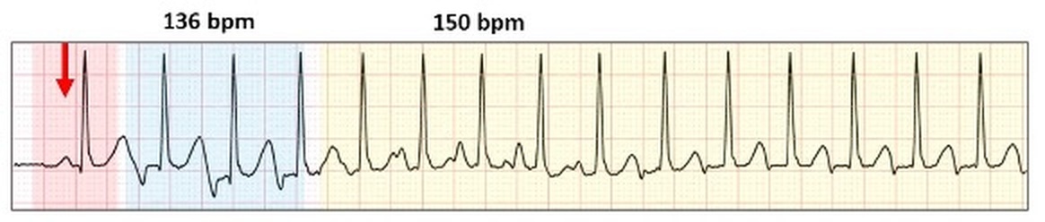
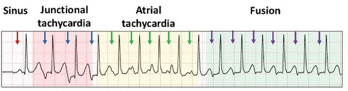
The first complex is sinus generated (red arrow and highlight). The tracing then deteriorates into a narrow complex tachycardia. Most physicians would call this a supraventricular tachycardia and leave it to the EP study to make a diagnosis. We need to look further.
There are two different tachycardia rates with different P wave configurations.
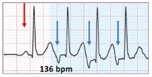
Let us review the first one (blue highlight).
Inverted P waves (blue arrows). This is most likely a junctional tachycardia.
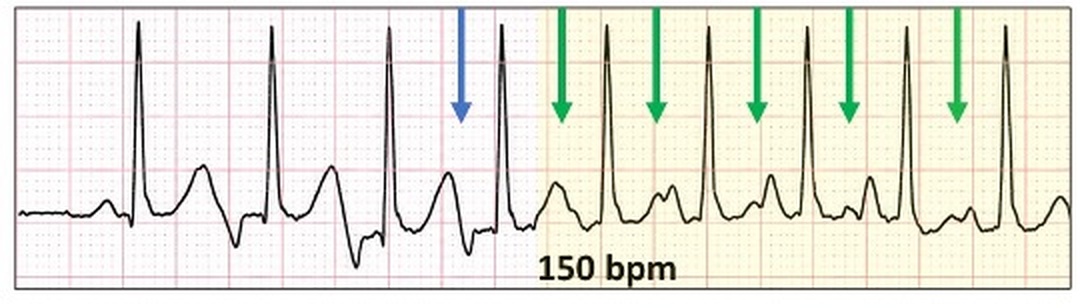
What about the second (yellow highlight)?
Transition to a tachycardia with P waves similar to the sinus P waves (green arrows). At 150 bpm, this is most likely an atrial tachycardia but also think atrial flutter.
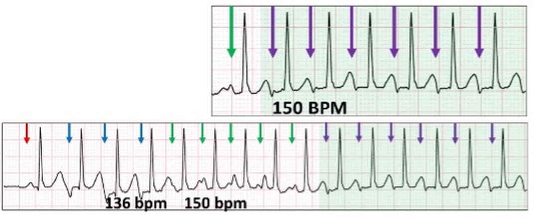
We have two supraventricular tachyarrhythmias competing with each other for ventricular depolarization. What happens when the rates approximate each other?
Once again, the P waves are inverted but smaller than earlier (green highlight). Both supraventricular ectopic foci depolarize the atria as fusion beats although probably only the junctional focus depolarizes the ventricle.
In summary:
This ECG highlights a poorly recognised and understood feature of junctional rhythm. If with junctional tachycardia, there is no retrograde conduction, then the atrium continues to depolarize separately. This can be sinus rhythm, an atrial tachyarrhythmia and even atrial fibrillation. However, with retrograde conduction, there is now competition between the supra-nodal and junctional foci as seen with our case study which resulted in fusion beats.
If you got it correct, please write to me.
What about the Schamroth ECG?
Firstly, wave the magic wand of AI.
This is the first description of sino-atrial Wenckebach block. Note the two dropped P waves (ghosted red arrows).
In 1970, I commenced work as a “novice” cardiologist at The Royal Melbourne Hospital. Training at that time was learning how to recognise the diastolic murmur of mitral stenosis and making sure the ECG and chest x-rays were the correct way up. As this was prior to coronary artery surgery, stenting and echocardiography, the ECG was a highly respected tool for the diagnosis of many conditions. I was fortunate to befriend a number of the gurus of ECG interpretation who taught me to look into the soul of an ECG. Today, even though the ECG is the first investigation performed by a cardiologist, the art of ECG interpretation has been lost because we depend on the new tools for cardiac diagnosis.
Dr Thura Thant asked for an opinion on this confusing ECG. By using arrows and highlight, the ECG bared its soul and gave me an answer. See if you can work it out? What do you think?
Fusion beats are an amalgam of two competing rhythms within the same chamber, atrial or ventricular. They are not “abnormal” beats and are a lesson in timing. For fusion beats to occur, two foci must commence propagation at or near the same time and both contribute to chamber depolarization. Both must be either atrial or ventricular and depending on the contribution of each, the resultant progeny have similarities to one or both parents.

.png)