To receive a free PDF copy of The Fundamentals of Electrocardiograph Interpretation by Harry Mond, subscribe to his email blog by entering your email address below.
Thank you! Your submission has been received!
Oops! Something went wrong while submitting the form.
Purchase the textbook
Purchase a hard cover or paperback copy of The Fundamentals of Electrocardiograph Interpretation by Harry Mond on Amazon.
Fusion beats are an amalgam of two competing rhythms within the same chamber, atrial or ventricular. They are not “abnormal” beats and are a lesson in timing.
For fusion beats to occur, two foci must commence propagation at or near the same time and both contribute to chamber depolarization. Both must be either atrial or ventricular and depending on the contribution of each, the resultant progeny have similarities to one or both parents.
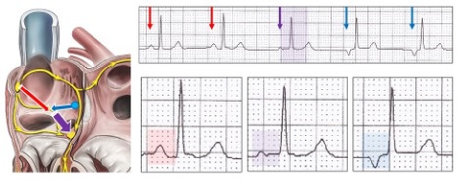
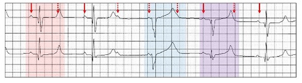
Atrial fusion beats occur as a result of competition between two atrial sites, usually sinus with ectopic atrial rhythm or atrial pacing. Only P waves show fusion.
Left: Two competing foci in the atrium, sinus (red arrow) and ectopic atrial (blue arrow) each depolarize a section of the atria (purple arrow). Above/ right: Sinus rhythm (red arrows) and ectopic atrial rhythm (blue arrows) with an atrial fusion beat and an identical QRS (purple arrow and highlight). Below/ right: Examples of the P wave; sinus (red highlight), ectopic atrial (blue highlight) and the resultant fusion beat (purple highlight), which is an amalgam of the other two P waves.
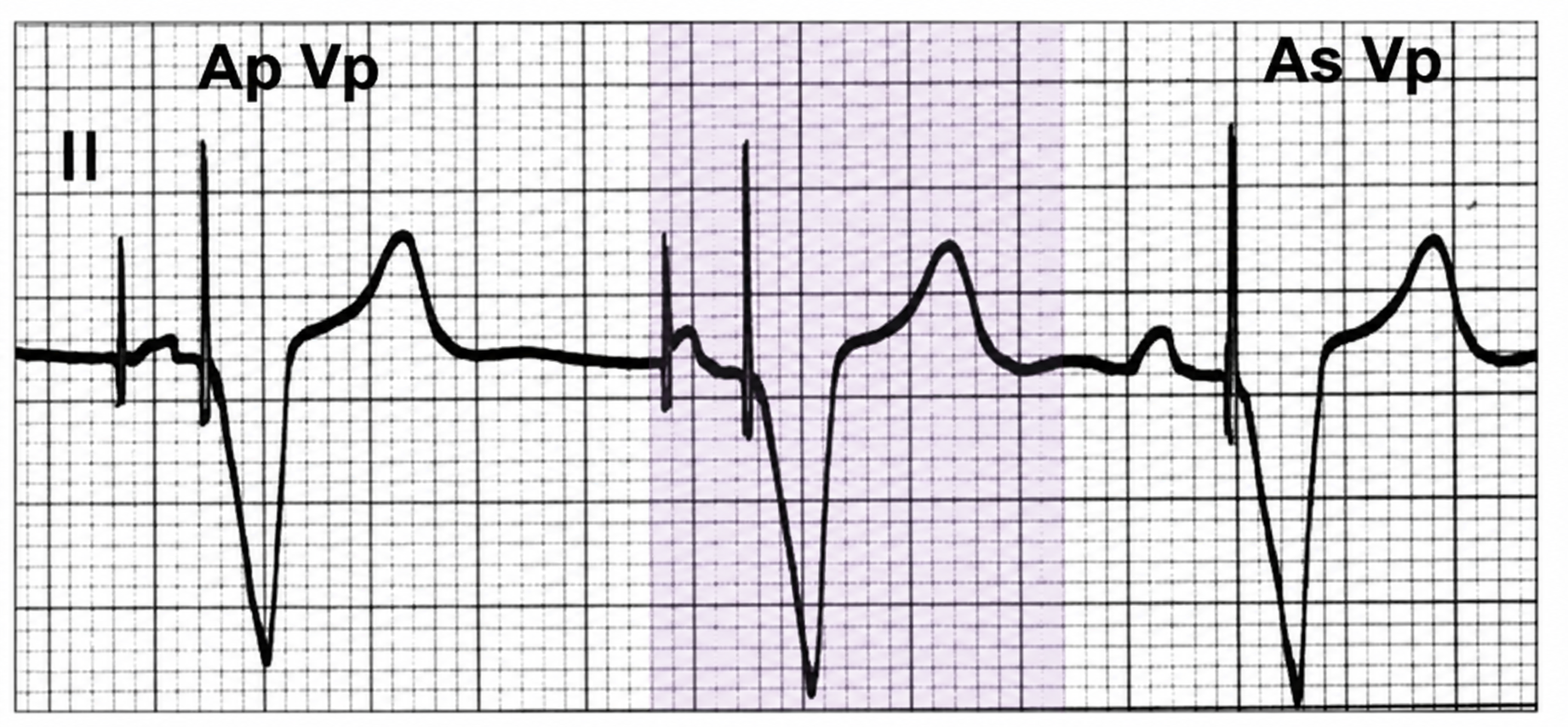
Dual chamber pacing There is both atrial pacing (Ap) and atrial sensing (As) and between lies atrial fusion (purple highlight) where the sinus node has commences depolarization of the atrium but is not sensed by the pacing lead. This is not pacemaker malfunction.
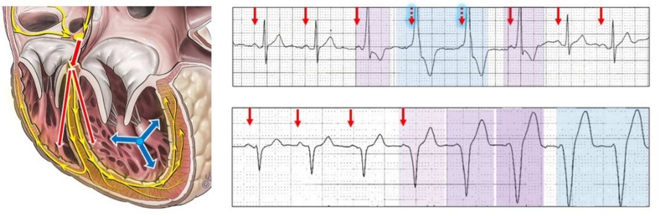
Ventricular fusion beats occur as a result of competition between two foci: usually one is sinus generated together with an ectopic ventricular focus or ventricular pacing. Only QRS complexes show fusion.
Left: Two competing rhythms in the ventricle, sinus with AV conduction (red arrows) and ventricular (blue arrows) each depolarize a section of the ventricle. Above/ right: Sinus rhythm (red arrows) with a four beat idioventricular run. There are ventricular fusion beats (purple highlight) on each side of the ventricular complexes (blue highlight). Each has a sinus P wave with a short PR interval preceding the fused QRS confirming fusion. Below/ right: Sinus rhythm (red arrows) with a continuing run of idioventricular rhythm (blue highlight) preceded by three ventricular fusion beats (purple highlight) gradually having an increased ventricular contribution.
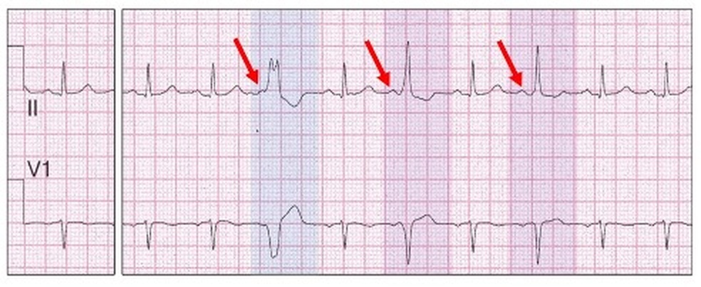
Late ventricular or “end-diastolic” ectopics, which occur in the PR interval of the next sinus cycle may fuse with the next sinus generated QRS producing a ventricular fusion beat.
End-diastolic ventricular bigeminy with a rapid sinus rhythm of 95 bpm. Each of the ventricular ectopics are 20 to 40 ms later into the PR interval as seen by the relationship to the preceding P wave (red arrows). The first ventricular ectopic (blue highlight) is not a fusion beat, whereas the next two (purple highlight) are. The gradual dominance of the sinus generated QRS results as the end-diastolic ectopic timing becomes later.
Ventricular fusion is also seen with intermittent complete AV block and competition with the ectopic ventricular escape focus when both are similar rates.
2:1 second degree AV block with a narrow conducted QRS (red highlight). When AV conduction completely fails the escape ectopic ventricular focus (blue highlight) has a near identical rate to the conducted native rhythm resulting in ventricular fusion beats (purple highlight).
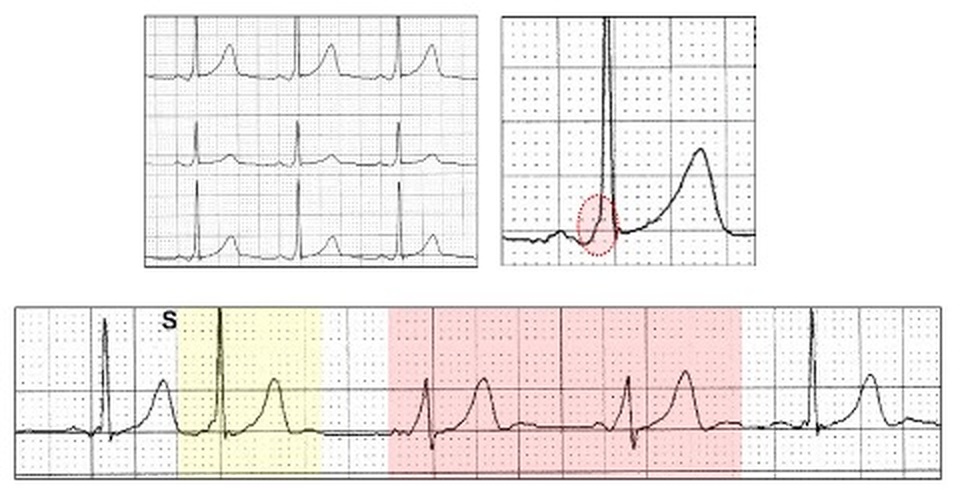
Ventricular fusion can also occur with the Wolff-Parkinson-White (WPW) ECG pattern of pre-excitation. Whereas the accessory pathway usually conducts to the ventricle faster than AV conduction resulting in a short PR interval, delta wave and broad QRS, on occasion AV conduction also occurs resulting in ventricular fusion. This results in a modified appearance to the WPW pattern.
Above: WPW pattern with a short but not abnormal PR interval (120 ms), small delta wave (red oval) and narrow QRS. Below: Following an atrial ectopic with the same features (yellow highlight), the conduction dynamics alter and the next two sinus complexes demonstrate a more complete WPW pattern (red highlight).
Ventricular fusion is frequently seen with ventricular parasystole and aids in the diagnosis.
Remind me about parasystole!
A parasystolic focus is an ectopic focus that competes with a primary (dominant) focus, so there are two intrinsic pacemakers each having different cycle lengths. It can occur at all levels in the heart and there is an entrance block protecting the secondary parasystolic focus, so that impulses cannot enter and reset the timing of the parasystolic focus. The parasystolic block is therefore unidirectional and whenever the parasystolic focus depolarizes, it will conduct to the surrounding myocardium, provided it is not refractory. Think of a pacemaker, programmed asynchronous (AOO, VOO) at a rate of about 40 bpm in competition with the dominant rhythm. The reported repetition rates for parasystole range from about 15 to 70 bpm with the slower rates being in the ventricle. On the ECG, at least three parasystolic complexes are required for the diagnosis, and long rhythm strips may be required.
Ventricular parasystole is the most easily and commonly recognised parasystole and usually occurs with sinus rhythm as the dominant rhythm. It masquerades as ventricular ectopy such as trigeminy or quadrigeminy but differs in that there are varying coupling intervals between sinus beats and the ventricular complexes. Because of an entrance block, the dominant rhythm cannot enter the parasystolic focus and reset its timing. Whenever the parasystolic focus depolarizes, it will conduct to the surrounding myocardium, provided the tissue is not refractory.
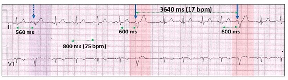
Sinus rhythm 75 bpm.
Unifocal, parasystolic ventricular ectopics (blue arrows, red highlight) with a fixed inter-ectopic interval of 3640 ms (17 bpm).
The ventricular parasystolic focus is not reset by the sinus generated QRS complexes and thus there is a variable sinus generated QRS to ventricular ectopic coupling interval (600 ms to 660 ms).
Ventricular fusion (purple highlight, blue stippled arrow) results when a scheduled ventricular parasystolic beat falls within the PR interval (end diastole) of the sinus complex.
Another example (purple highlight):
When the ectopic timing occurs during the ventricular refractory period, the parasystolic output is inhibited (green highlight).
By far the most common cause of ventricular fusion beats is with atrial fibrillation and ventricular pacing. There are usually frequent pauses with the native rhythm allowing ventricular pacing. At the end of the pause, a native ventricular complex may compete with ventricular pacing resulting in a fusion beat (purple highlight).
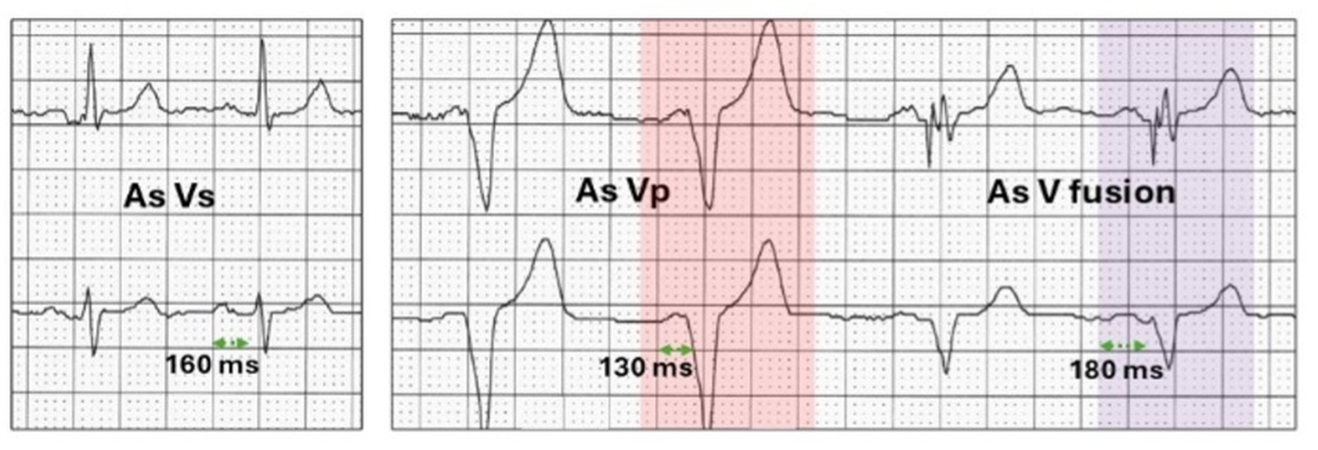
With dual chamber pacing, the PR interval of the native rhythm (As Vs) may be near identical to the programmed AV delay so that a sinus generated native ventricular wave of depolarization fuses with the paced QRS for long periods. The ventricular fusion is usually not recognized on the ECG and can be prevented by ventricular pacing minimization algorithms (section 16.17). They can also be recognized on Holter monitoring when “forced” ventricular pacing occurs during auto-threshold testing algorithms (section 16.18).
Medtronic dual chamber auto-threshold testing algorithm. With native rhythm (As Vs), the PR interval is 160 ms. During the test, the AV delay shortens to 130 ms with forced ventricular pacing (Vp, red highlight). At the end of the test, there are no visualized ventricular low voltage stimulus artefacts on the ECG, and the rhythm converts to the programmed AV delay of 180 ms with ventricular fusion (purple highlight).
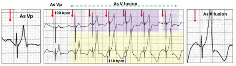
When ventricular fusion occurs with biventricular pacing, there are now three ventricular waves of depolarization involved in the fused complex.
Left: Sinus rhythm (As, red arrow) with biventricular ventricular pacing (Vp).Right: run of ventricular tachycardia 110 bpm (yellow highlight) marginally faster than the sinus rate 100 bpm, (red arrows). The ventricular stimulus artefact continue through the ventricular tachycardia resulting in ventricular fusion. The fusion commences as predominantly paced rhythm (light purple highlight) and becomes more progressively tachycardia (deepening purple highlight) and at the end there is once again increasing pacing contribution.
Retrograde conduction is defined as conduction from an ectopic site to the atrium via the AV node or accessory pathway and referred to as VA conduction. II results in a P wave configuration which differs from an anterograde P wave, and in most ECG leads is inverted. The site from where the impulse originates can be the AV node, Bundle of His, distal conducting system or the body of the ventricles including ventricular pacing. On the ECG, the QRS complex usually precedes the inverted retrograde P wave but on occasion may follow it.