To receive a free PDF copy of The Fundamentals of Electrocardiograph Interpretation by Harry Mond, subscribe to his email blog by entering your email address below.
Thank you! Your submission has been received!
Oops! Something went wrong while submitting the form.
Purchase the textbook
Purchase a hard cover or paperback copy of The Fundamentals of Electrocardiograph Interpretation by Harry Mond on Amazon.
In our last “what do you think?”, we reviewed the ventricular ectopic and the range of “geminy” groupings. The above ECG is an extension of this but before we can proceed to analyse the tracing, we need to revisit once again the ventricular ectopic, but this time, the compensatory pause.
The compensatory pause extends from the commencement (or maybe the end) of the ectopic QRS/T, until the onset of the next P wave.
There are two types of compensatory pauses, fulland partial.
I have never read an explanation of these, so I have created my own.
Full compensatory pause.
Sinus rhythm (red vertical lines). There is a concealed, non-conducted sinus P wave (red stippled vertical line) buried in the ectopic QRS. Following is a pause until the next sinus cycle. This is called a full compensatory pause. There is no resetting of the sinus cycle and hence the timing of two sinus cycles with the embedded ventricular ectopic is identical to two sinus cycles without an embedded ventricular ectopic (1700 ms).
Partial compensatory pause.
Sinus rhythm (red vertical lines) rate 60 bpm (1000 ms). There is a ventricular ectopic and embedded within is a premature P wave (blue stippled vertical line), which results from retrograde conduction to the atria. As this precedes the next sinus P wave by 200 ms, the next sinus P wave is inhibited, and the sinus cycle is reset. The next sinus P wave then occurs in 1000 ms. The two sinus cycles with the embedded ventricular ectopic occur 1800 ms apart, whilst two sinus cycles without the embedded ventricular ectopic are 2000 ms. Because it is shorter, it is referred to as a partial compensatory pause.
Note that even though the partial compensatory pause is “shorter” than the full compensatory pause, it nevertheless, doesn’t look short on the ECG,
Depending on the sinus rate, prematurity, and width of the ventricular ectopic, the embedded non-conducted sinus P wave can sometimes appear after the ectopic QRS or T wave and is now not concealed.
Sinus rhythm (vertical lines)with two early ventricular ectopics in a bigeminal sequence. At the termination of the ectopic QRS in the ST segment, the non-conducted P wave (red highlight, green vertical line) can be seen, and the sinus cycle is not interrupted.
If the timing is appropriate, this P wave can conduct to the ventricle resulting in an interpolated ventricular ectopic, albeit with a prolonged PR interval, as the AV junction is still partially refractory.
Sinus rhythm, (vertical redlines) with a PR interval of 200 ms (yellow highlight). Following the ventricular ectopic, the PR interval is extended to 400 ms (blue highlight),thus increasing the R-R interval from 1140 ms to 1340 ms. There is NO concealed nor non-conducted sinus P wave and NO compensatory pause. An interpolated ventricular ectopic may on occasion not extend the PR interval or the P wave is concealed in the ectopic.
Another scenario occasionally seen is very marked extension of the PR interval (blue highlight).
This results in an illusionary or pseudo-compensatorypause ( red stippled arrow), with the P wave near the middle of the pause.
Remember the complexity of the timing may also result in aberrant ventricular conduction of the next sinus beat.
Sinus rhythm (red vertical lines), and a ventricular ectopic (red highlight) with the next P wave concealed in the ectopic T wave (blue vertical line), but conducts to the ventricle, with aberrancy (yellow highlight).
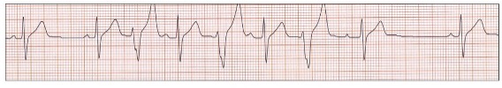
Let us return to our ECG:
Sinus rhythm 60 bpm (red vertical lines) with a normal PR interval (yellow highlight) and interpolated ventricular bigeminy (red highlight) with PR extension (blue highlight).
and interpolated ventricular quadrigeminy (red highlight).
Did someone say, “what about interpolated ventricular couplets?”
Timing is critical in allowing two ectopics in bigeminy to fall into a non-refractory zone and allow AV conduction, thus maintaining conduction of all sinus beats.
Sinus rhythm (red vertical lines) with a normal PR interval (yellow highlight) and a ventricular couplet (red highlight). The first ventricular ectopic in the couplet falls late in AV conduction. The P wave of the next sinus beat is concealed, and the PR interval extended (blue highlight), so there are no non-conducted sinus beats and the couplet interpolated.
Can a triplet be interpolated?
No P waves are seen, and we must assume sinus bradycardia (red vertical lines). There is one ( or two)non-conducted sinus P waves in the body of the triplet but no compensatory pause making this rhythm interpolated.
No tricks. Just a selection of tracings from a Holter study.Look at each one carefully, use calipers, arrows and highlight and write down your conclusions.