To receive a free PDF copy of The Fundamentals of Electrocardiograph Interpretation by Harry Mond, subscribe to his email blog by entering your email address below.
Thank you! Your submission has been received!
Oops! Something went wrong while submitting the form.
Purchase the textbook
Purchase a hard cover or paperback copy of The Fundamentals of Electrocardiograph Interpretation by Harry Mond on Amazon.
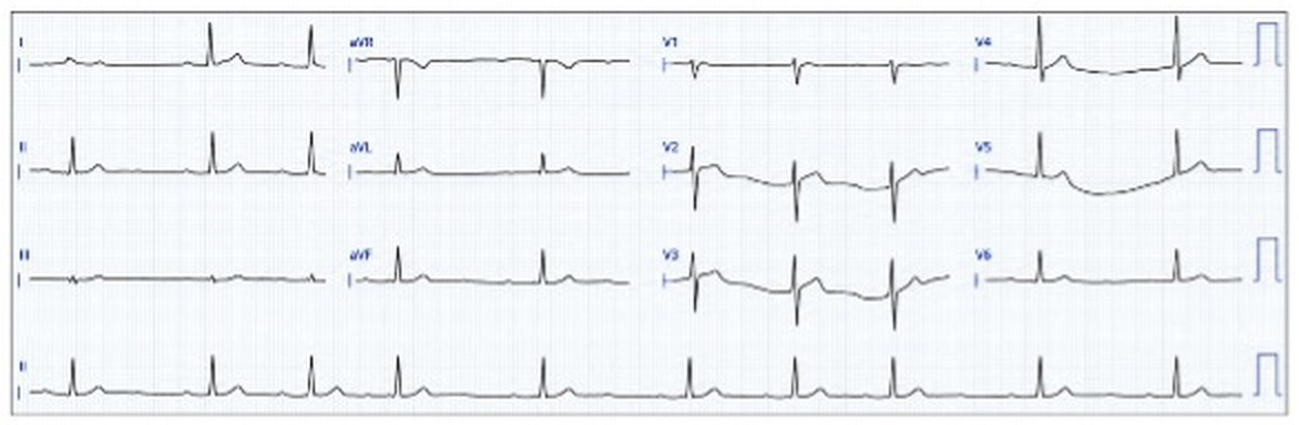
Dr Thura Thant asked for an opinion on this confusing ECG.
By using arrows and highlight, the ECG bared its soul and gave me an answer.
See if you can work it out?
What do you think?
We need to use the rhythm strip.
There is a pattern with the QRS complexes.
Repeated three QRS complexes (red highlight) at a rate of 75 bpm, followed by a short pause of 1200 ms (50 bpm).
Prior to each QRS is a P wave (red arrows) which we assume are sinus generated.
When we see group beating with a pause, think Wenckebach.
However, sino-atrial Wenckebach would have P-P shortening and the pause a multiple of the P-P interval. This doesn’t fit!
Type II sino-atrial block would have a longer pause and again a multiple of the P-P interval.
We need to look harder.
Are there dropped beats?
There appear to be scattered dropped P waves.
Do these P waves align with the other P waves?
The non-conducted P waves (large red stippled arrows) align with the conducted P waves in that they follow a conducted P wave after 800 ms, which is the P-P interval. However, following the non-conducted P waves the next conducted P wave occurs after 400 ms (150 bpm) or half the P-P interval.
This doesn’t make sense unless the atrial rate is 150 bpm (an atrial tachyarrhythmia).
Atrial flutter with 2:1 AV block is ruled out so this is an atrial tachycardia (red and blue arrows).
Atrial tachycardia (blue arrows).
The ventricular rate is controlled and thus there is AV block.
Is this Wenckebach AV block?
There is a very minor increase in the PR interval (green stippled arrows) before the dropped beat (blue stippled arrow). There are alternating concealed non-conducted P waves buried in the T waves of the previous beat.
This is alternating Wenckebach AV block.
Alternating Wenckebach AV block
The AV junction, with its decremental conduction, plays a vital role in controlling the propagating wave of depolarization on its passage to the ventricle. Not only is the traffic slowed but under certain circumstances partial or complete blocks occur as can be seen with Wenckebach AV block. What is poorly understood is that more than one block can occur in series both physiologically with rapid atrial rhythms and pathologically with conduction system disease. When two blocks occur in series, it is called alternating Wenckebach AV block and because they occur at different levels, they are multilevel blocks.
Particularly with rapid atrial rhythms, alternating Wenckebach AV block is often difficult to visualize on the surface ECG and therefore the tracings are best described when associated with a bradyarrhythmia resultant from advanced degenerative changes in the AV junction. The tracings are usually mistaken for complete AV block with the difference being the presence of an irregular ventricular rate but with regular Wenckebach sequences.
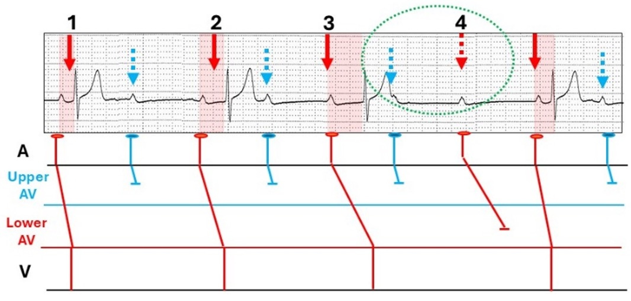
Sinus rhythm (arrows) with two blocks; one high (light blue) and the other low (red), with the combination, a multilevel block.
The positioning of the blocks vary and for a simplistic explanation, the conducting impulse is first subjected to 2:1 AV block (blue) probably within the AV node.
The alternating conducted impulses then encounter 4:3 Wenckebach (red) either in the AV node or more distal conducting system.
A new sequence commences without an intervening distal blocked beat.
With a slow sinus rate, only two consecutive non-conducted sinus P waves occur (green stippled open oval).
Can occur with a bundle branch block, syncope, and sudden death and thus it is very likely to be pathological and associated with conduction tissue disease.
With atrial tachyarrhythmias, alternating Wenckebach AV block protects the ventriclesfrom rapid atrial rates and is thus physiological but in the presence of atrial pathology. The arrhythmia can be easily identified when the P waves are discrete as in this very short run of atrial tachycardia diagnosed as an atrial couplet (SVE pair).
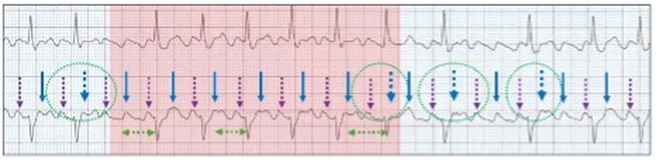
Sinus rhythm (red arrows), rate 56 bpm. Five beat run of rapid atrial tachycardia (188 bpm) with two conducted P waves (blue solid arrows) demonstrating 3:2 Wenckebach AV block; PR intervals 240 ms and 280 ms and a dropped beat (large stippled blue arrow). There are alternating non-conducted P waves (stippled pink arrows) and the run is terminated by two consecutive non-conducted P waves.
When the rate of the atrial tachyarrhythmia is relatively slow, the alternating 2:1 AV block may be sufficient to control the ventricular response so that the Wenckebach sequences may be very long and without dropped beats.
Slow atrial tachycardia (112 bpm) with non-completed alternating Wenckebach AV block. There is 2:1 AV block (purple stippled arrows) and alternating conducted beats (blue arrows) with a gradual increase in the PR interval and no dropped beats. The ventricular response is slow and near regular.
When confronted with Wenckebach sequences and a possible atrial tachyarrhythmia, the clue to alternating Wenckebach AV block lies in the short pause. Look for the P wave(s) and are the sequences correctly linked.
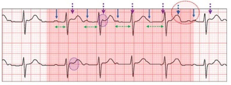
Wenckebach sequence (green stippled arrows, red highlight). Following the dropped beat (blue thick stippled arrow), the next sequence occurs with the P wave timing shortened (red stippled open oval). There is ST distortion (purple stippled open ovals) which line up with non-conducted P waves (purple stippled arrows). The diagnosis is atrial tachycardia, 190 bpm, and alternating Wenckebach AV block.
Most cases of alternating Wenckebach AV block occur with atrial flutter or a macro-reentrant atrial tachycardia and are frequently missed on the surface ECG because of poorly visualized P waves. Like conventional Wenckebach AV block, there are sequences separated by short pauses.
Here are two examples with prominent P waves.
Atrial flutter (arrows), rate 250 bpm with a sequences of six ventricular responses (red highlight), each with an increasing PR interval (green stippled arrows) until a dropped beat (wide blue stippled arrow). There are alternate non-conducting P waves (purple stippled arrows) and the sequence is terminated by two dropped beats (green stippled open ovals). Other sequences have three dropped beats associated with a short pause.
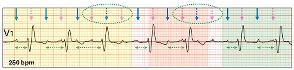
Atrial flutter rate 250 bpm with a controlled irregular ventricular response. There are three sequences. The first (yellow highlight) is 4:3 Wenckebach AV block (blue arrows) alternating with non-conducted flutter waves (pink stippled arrows) and three consecutive dropped P waves (green open stippled oval). Then follows two more sequences. Note the conducted P wave is not necessarily the closest to the QRS.
Physiologic alternating AV Wenckebach is an amazing rate control system protecting the ventricle from rapid atrial arrhythmias. AV node conduction can skip from any combination of Wenckebach AV block and coupled with 2:1 AV block varies the number of dropped P waves to fine control the ventricular response. With atrial flutter having 3 or 4:1 AV block, the surface ECG gives no clues as to the number of multilevel blocks involved which are believed to lie at several levels in the AV node and His bundle.
Let us return to our case study.
In summary:
Although the atrial tachycardia represents atrial pathology, the alternating Wenckebach response at the AV node is a physiologic protective mechanism.
Dr Thura Thant asked for an opinion on this confusing ECG. By using arrows and highlight, the ECG bared its soul and gave me an answer. See if you can work it out? What do you think?
Fusion beats are an amalgam of two competing rhythms within the same chamber, atrial or ventricular. They are not “abnormal” beats and are a lesson in timing. For fusion beats to occur, two foci must commence propagation at or near the same time and both contribute to chamber depolarization. Both must be either atrial or ventricular and depending on the contribution of each, the resultant progeny have similarities to one or both parents.







.jpg)



.jpg)