To receive a free PDF copy of The Fundamentals of Electrocardiograph Interpretation by Harry Mond, subscribe to his email blog by entering your email address below.
Thank you! Your submission has been received!
Oops! Something went wrong while submitting the form.
Purchase the textbook
Purchase a hard cover or paperback copy of The Fundamentals of Electrocardiograph Interpretation by Harry Mond on Amazon.
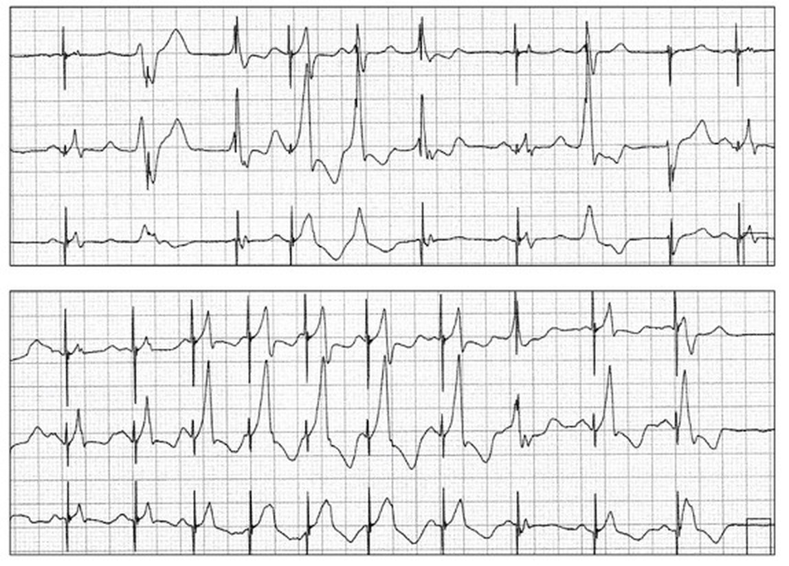
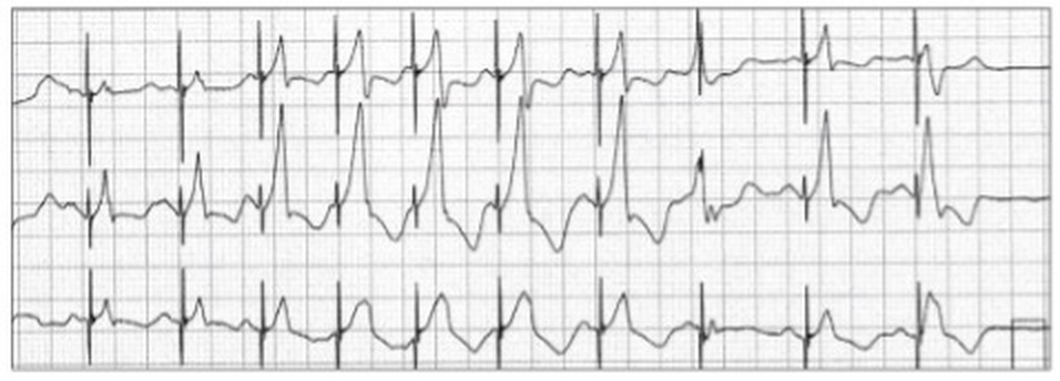
There are a number of different QRS complexes with associated stimulus artefacts. Let us review the conventional ones first.
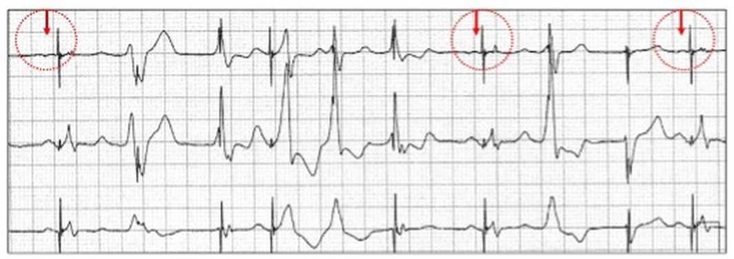
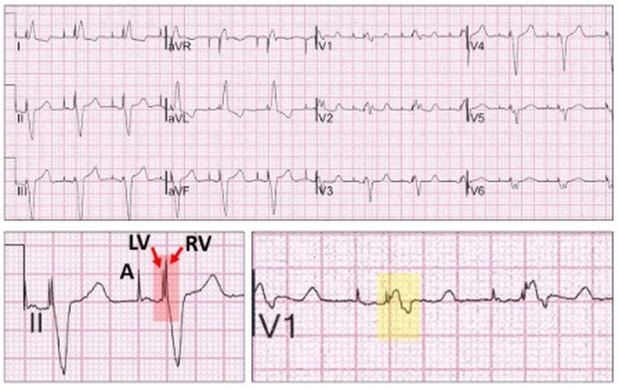
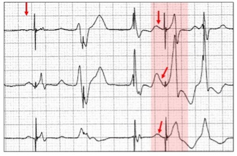
Three stimulus artefacts preceded by a small P wave (red arrow, open red circle).
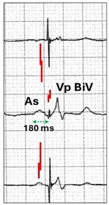
On careful inspection, the stimulus artefact in each lead is split into two, one following the other by about 40 ms. The “paced” QRS is also narrow.
These are features of biventricular pacing.
Biventricular pacing
A non-bradyarrhythmic indication for permanent cardiac pacing, is a form of dual-site pacing with near simultaneous depolarization of the right and left ventricles. It has been found useful for symptomatic patients with a non-ischaemic congestive cardiomyopathy on optimal tolerated medical therapy, New York Heart Association class 3 or 4 and a left bundle branch block on the ECG.
Having a left bundle branch block results in early septal depolarization and contraction, whilst the remainder of the left ventricle has late activation. Although in an otherwise normal heart this is usually asymptomatic, in one with left ventricular dysfunction from a congestive cardiomyopathy, the resultant ventricular dyssynchrony further reduces left ventricular function and remains uncorrected with conventional medical therapy. Simultaneous depolarization of the left and right ventricles may correct the dyssynchronous contraction, and this is referred to as cardiac resynchronization therapy (CRT).
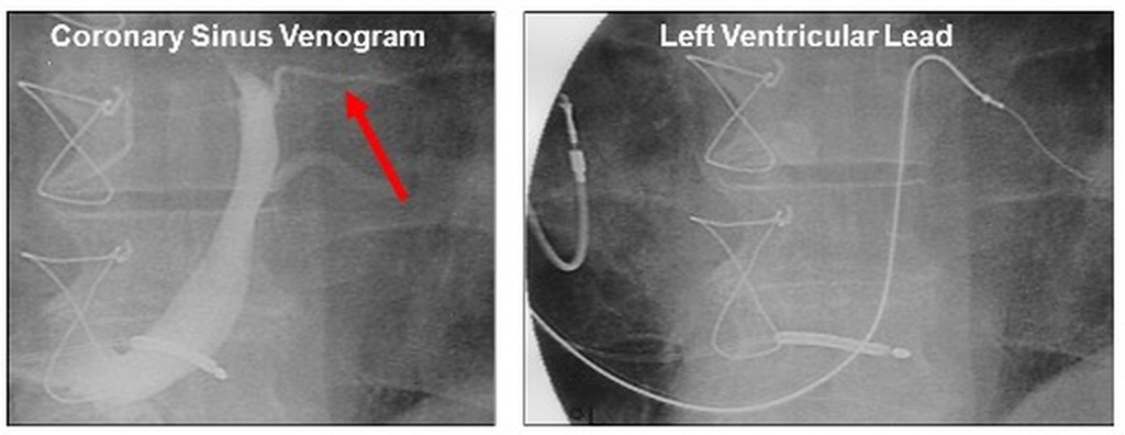
The transvenous implantation involves placement of an additional ventricular lead into a cardiac vein via the coronary sinus after a venogram to identify the vein (red arrow).
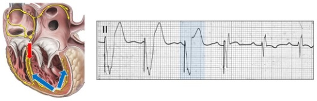
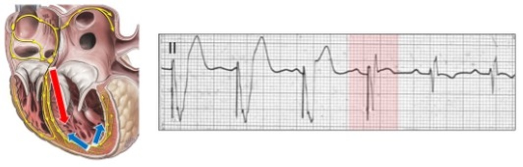
The left ventricle is usually paced immediately before the right to ensure optimal synchronization, with the V-V stimulus artefact timing programmable. This results in a right ventricular bundle branch block configuration (yellow highlight).
When the V-V stimulus artefact period between left (LV) and right (RV) stimulation is ≥ 40 ms, two stimulus artefacts can be identified on the ECG (red highlight). With dual chamber pacing, these follow atrial pacing (A) or sensing.
The appearance of the right bundle branch block configuration will vary considerably depending on the V-V stimulus artefact timing.
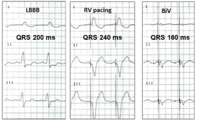
With no interval (0 ms), there is a left bundle branch block configuration and two fused stimulus artefacts. The two stimulus artefacts can be recognised if their vector (polarity) is different. With an 80 ms timing, the appearance is a right bundle branch block configuration with two now separated stimulus artefacts (LV, RV). Such a timing would not allow synchronization. With a 40 ms timing, the bundle branch block is a transition between right and left configurations.
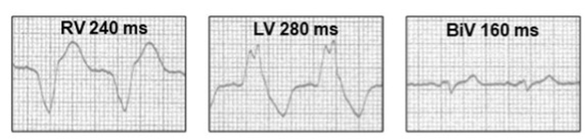
With biventricular pacing (BiV), there is usually QRS narrowing (160 ms) compared to the initial left bundle branch block (LBBB 200 ms) or single chamber right ventricular pacing (RV 240 ms).
Although single chamber left ventricular pacing (LV 280 ms) may also provide more physiologic pacing, the QRS duration is similar to right ventricular pacing (RV 240 ms), whereas biventricular pacing may have a much shorter duration (BiV 160 ms).
Let us return to our case study.
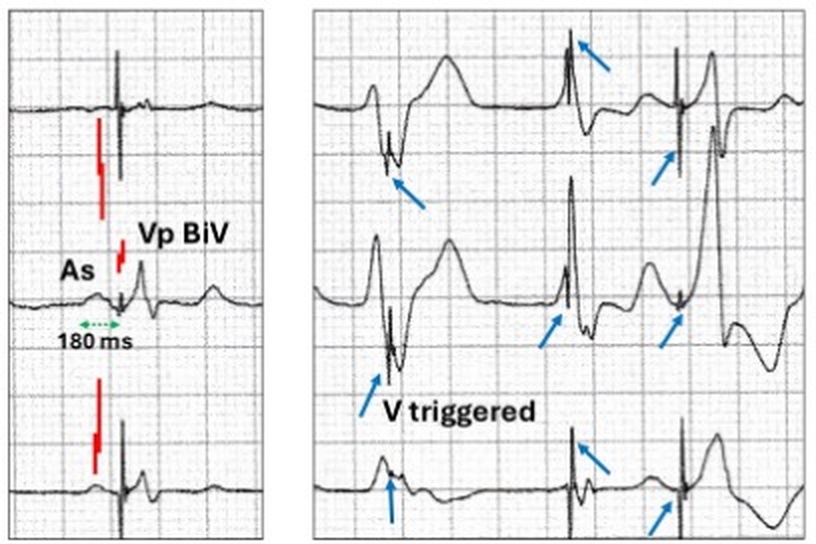
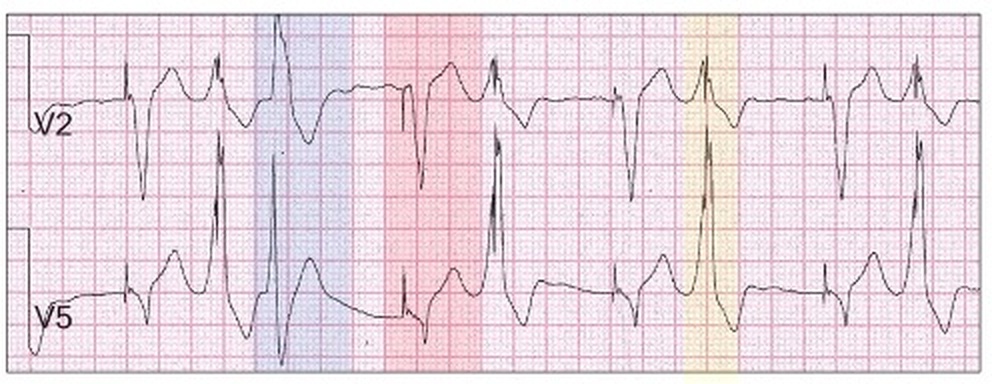
There are other broad QRS complexes with varying configurations which have stimulus artefacts (blue arrows) within the body of the QRS, rather than at the commencement.
This is ventricular triggered (VVT) pacing.
Ventricular triggered (VVT) pacing.
Early unipolar VVI sensing circuits were subject to oversensing and could lead to symptomatic pacing inhibition from sensed artefact or skeletal myopotentials.
An early non-programmable unipolar VVI pacemaker implanted in the abdomen. Sitting from the prone position resulted in pre-syncope as a result of abdominal skeletal myopotential inhibition. As this was pre-programmability, the pulse generator was surgically replaced. Consequently, prior to programmability, most of the early synchronous or demand pacemakers were VVT.
With VVT pacing, the full energy of the pacing impulse is delivered into the sensed intrinsic QRS (yellow highlight) resulting in a bizarre appearance with a premature stimulus artefact embedded within the intrinsic QRS at the exact moment of sensing.
If there are no sensed intrinsic beats, pacing appears normal (red highlight). The energy delivered into intrinsic beats usually does not contribute to the QRS, is premature and thus the pacing rate appears to increase and is energy expensive. There is also a very premature ectopic as part of a couplet (blue highlight) which does not have embedded stimulus artefacts to be explained later.
What do these embedded timulus artefacts have to do with biventricular pacing?
The objective of biventricular pacing is to pace the ventricle greater than 90% of the time, thus optimizing resynchronization. However, with atrial fibrillation or frequent ventricular ectopy, this may become a challenge. A novel, but unproven pacemaker algorithm is to attempt to pace both ventricles on sensing of the native ventricular rhythm. This is essentially ventricular triggered pacing (VVT or more accurately DDT). It is present as a programmable function in at least two manufacturer’s biventricular pulse generators. Ventricular sensing of the native rhythm is variable and may be late and therefore biventricular pacing is unlikely to contribute significantly to ventricular depolarization and hence resynchronization. With frequent native rhythm, triggered biventricular pacing is energy expensive and its value unproven.
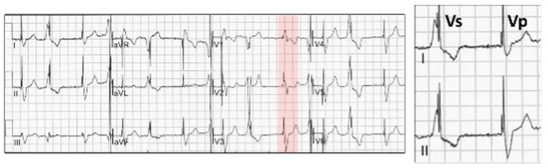
Biventricular pacing with a right bundle branch block configuration (red highlight) with two stimulus artefacts (Vp), the first left ventricular and 40 ms later right ventricular. There is ventricular bigeminy (Vs) with the two stimulus artefacts embedded late within the native QRS, suggesting that meaningful ventricular resynchronization is unlikely.
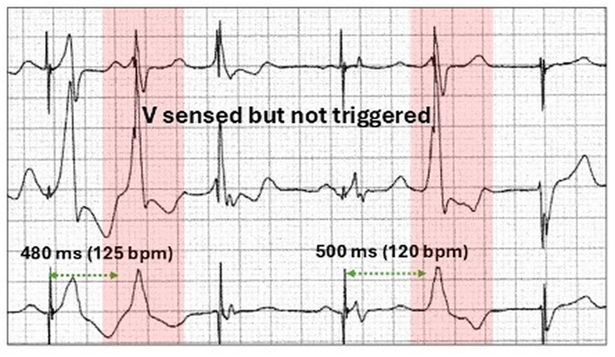
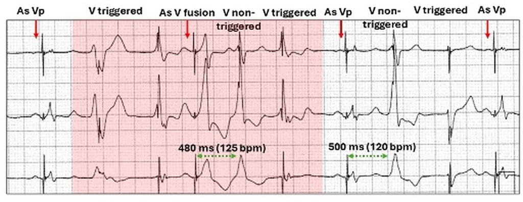
As seen in an earlier illustration, not all native complexes have embedded stimulus artefacts. In our case study, there are two premature broad QRS complexes (red highlight) without embedded stimulus artefacts, although there is otherwise appropriate ventricular sensing.
Both ectopic QRS complexes are very premature with short coupling intervals at 480 and 500 ms.
How do we explain this?
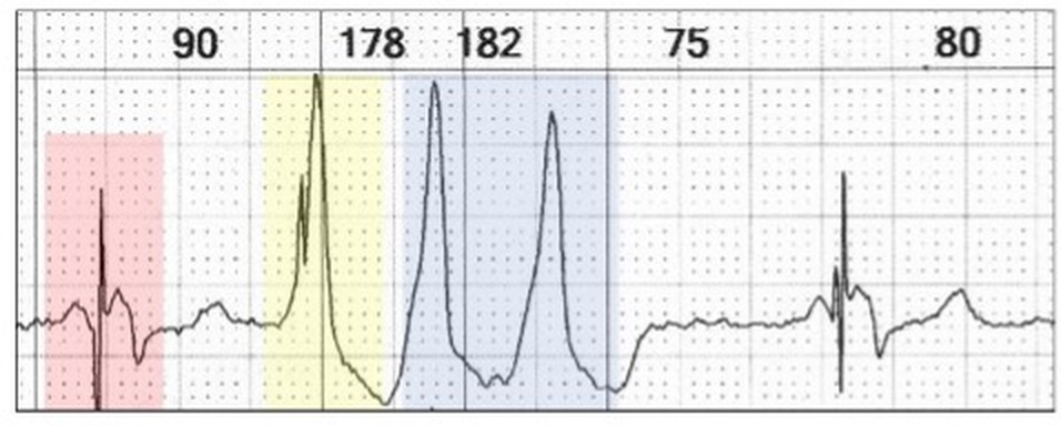
With ventricular triggered pacing, there is concern regarding oversensing of electrical interference resulting in rapid ventricular pacing. Consequently built into all triggered systems is a programmable upper rate limit with the default being about 120 to 130 bpm.
Triggered single chamber biventricular pacing with two stimulus artefacts, each with an opposite vector (red highlight). A ventricular triplet is sensed as suggested by the timing of the next paced beat, The first ventricular ectopic demonstrates triggering with embedded stimulus artefacts (yellow highlight), whereas the next two (blue highlight) do not trigger an output as the upper rate exceeds ~130 bpm (178 bpm, 182 bpm).
Thus the absence of stimulus artefacts embedded into sensed QRS complexes is not abnormal pacemaker function but related to ventricular rates exceeding the upper rate limit for sensing.
There is another broad premature QRS but with a preceding stimulus artefact.
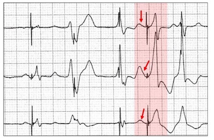
Broad premature QRS (red highlight) with a stimulus artefact at the commencement of the QRS. Although very subtle, there is a faint notching on the downslope of the preceding T wave, suggesting a concealed sinus P wave.
The broad QRS is a fusion beat between the sinus conducted biventricular depolarization and a ventricular ectopic.
Paced ventricular fusion beats.
Fusion beats occur whenever there are at least two foci in either the atrium or ventricle competing against each other and represent a lesson in timing. They are the amalgam of either two rhythms or late ectopy within the same chamber and are common when a demand pacemaker competes usually with sinus rhythm. Both are responsible for partial depolarization of the respective chambers and depending on the contribution of each, result in progeny with similarities to one or both parents. There is failure by the pacemaker to sense the spontaneous beat as it has not depolarized sufficient amount of ventricular myocardium to allow sensing. Although there is apparent sensing failure, this is not pacemaker malfunction.
A ventricular pacing focus competes with sinus generated beats conducted via the His-Purkinje system.
Ventricular pacing (VVI) with the fusion of a sinus generated beat (red arrow) with a predominantly ventricular paced beat (blue highlight, blue arrows).
The next sinus generated QRS is a few ms earlier (red arrow) and now the fusion beat is predominantly sinus generated (red highlight).
With dual chamber pacing, it is possible for the sinus generated paced QRS (DDD) to compete with a late (end-diastolic) ventricular ectopic resulting in a fusion beat. Another situation is fusion between a conducted sinus beat and sinus triggered ventricular pacing (VDD, DDD) so that the sinus P wave generates both AV conduction and ventricular pacing. This is surprisingly common with sinus node disease. The intrinsic AV conduction time is similar to the programmed AV delay, so that the conducted beat and ventricular pacing contribute to the generated QRS. As this is energy wasteful and may lead to left ventricular dysfunction, the AV delay should be programmed longer or a ventricular pacing minimization algorithm programmed ON (section 16.17).
Let us return to our case study.
Because the sinus generated biventricular QRS complexes are small and narrow in comparison to the ventricular ectopic, the morphology of the resultant fusion beat is dominated by the ectopic. This is actually an amalgam of three generated QRS complexes, two from a pacemaker activation and one from an ectopic ventricular focus.
The fusion beat is highlighted in red. There is small P wave prior to it embedded in the downstroke of the wave creating a small notch (red arrows).
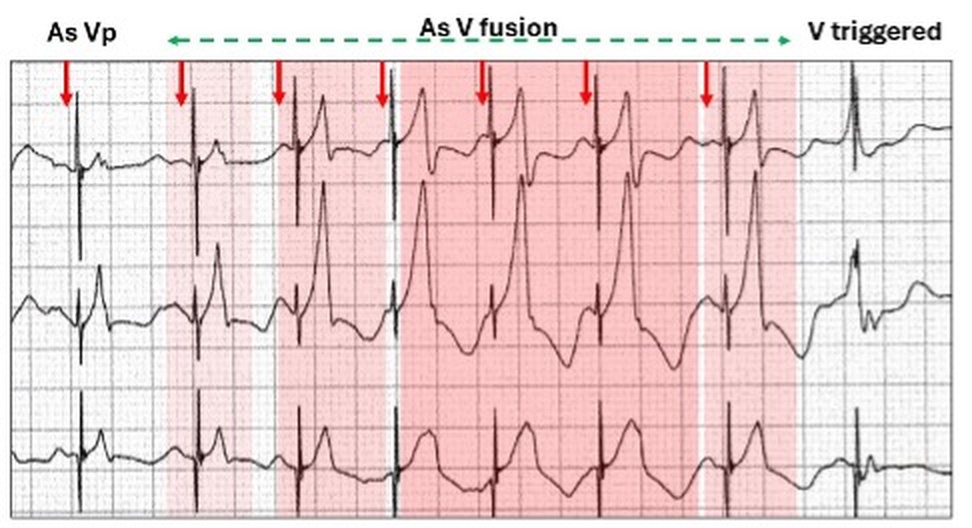
The second tracing in our case study was more difficult to interpret but highlights the differing contributions of the parents to the fused QRS complexes.
Every complex in the tracing has a preceding or embedded stimulus artefact.
Following the first complex (As Vp), there is a run of competition between biventricular pacing and ventricular tachycardia. The timing has created a run of ventricular fusion, which commences with predominant biventricular contribution (light red highlight) and then increasing ectopic contribution (deepening red highlight) and finally more biventricular contribution as the P wave reemerge.
In summary:
Sinus rhythm/tachycardia with biventricular pacing (As VP, red arrows)
Multifocal ventricular runs (red highlight) with:
Ventricular triggering.
Premature ventricular complexes in the pacemaker triggering refractory period.
Fused biventricular pacing and ventricular complexes.