To receive a free PDF copy of The Fundamentals of Electrocardiograph Interpretation by Harry Mond, subscribe to his email blog by entering your email address below.
Thank you! Your submission has been received!
Oops! Something went wrong while submitting the form.
Purchase the textbook
Purchase a hard cover or paperback copy of The Fundamentals of Electrocardiograph Interpretation by Harry Mond on Amazon.
Wandering atrial pacemaker or“chaotic atrial mechanism” usually refers to sinus rhythm with at least two ectopic atrial foci. When the rate is greater than 100 bpm, it is called multifocal atrial tachycardia. This is usually in patients with chronic airways disease, is a precursor to atrial fibrillation and has a poor prognosis.
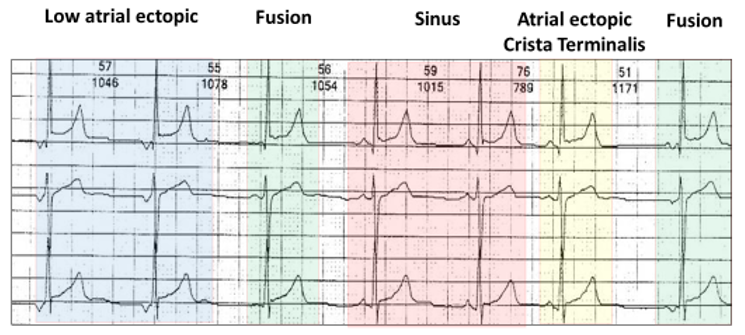
Here is an example of a wandering atrial pacemaker:
Two sinus beats (red highlight). One complex with a P wave similar to the sinus P wave, but premature and probably originates from an area within the crista terminalis, adjacent to the sinus node (yellow highlight). There is also a low atrial couplet(blue highlight). Two of the beats are not premature and have different P wave morphologies and are likely to be fusion beats (green highlights).
Atrial fusion is there for every important in determining if it is a wandering atrial pacemaker. For this reason, wandering atrial pacemaker is frequently misdiagnosed.
Let us return to the ECG
The first two complexes are sinus (red highlight). The last two beats are low atrial ectopics or more accurately, escape rhythm. The middle complex is an amalgam between the sinus and low atrial beats and is therefore an atrial fusion beat.
The rhythm is not wandering atrial pacemaker as there is only one atrial ectopic focus.
The diagnosis is low arterial escape rhythm and atrial fusion.
Retrograde conduction is defined as conduction from an ectopic site to the atrium via the AV node or accessory pathway and referred to as VA conduction. II results in a P wave configuration which differs from an anterograde P wave, and in most ECG leads is inverted. The site from where the impulse originates can be the AV node, Bundle of His, distal conducting system or the body of the ventricles including ventricular pacing. On the ECG, the QRS complex usually precedes the inverted retrograde P wave but on occasion may follow it.