To receive a free PDF copy of The Fundamentals of Electrocardiograph Interpretation by Harry Mond, subscribe to his email blog by entering your email address below.
Thank you! Your submission has been received!
Oops! Something went wrong while submitting the form.
Purchase the textbook
Purchase a hard cover or paperback copy of The Fundamentals of Electrocardiograph Interpretation by Harry Mond on Amazon.
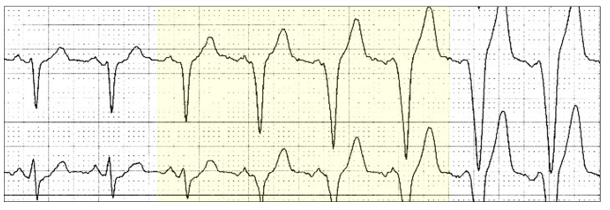
To the left is unipolar ventricular pacing (VVI) and to the right, sinus rhythm with first degree AV and bundle branch block. Lying between these are two different QRS configurations referred to as ventricular fusion beats.
Fusion beats are an amalgam of two competing rhythms. Both are responsible for partial depolarization of the respective chambers and depending on the contribution of each, result in progeny with similarities to one or both parents.
In the above example, the fusion beat to the left (red highlight) looks similar to the ventricular paced rhythm and the one to the right (yellow highlight), looks similar to sinus rhythm.
Let me explain this in more visual detail.
The fusion complex that looks like the ventricular pacing parent (red highlight) is generated predominantly from pacing with the endocardial transvenous lead tip in the right ventricle. With pacing, there is a set cycle length and if the lead does not sense native ventricular depolarization (VVI), it will then pace the heart (red arrows).However, a sinus beat may have already depolarized the atrium (P wave, blue vertical arrow) and progressing through the conducting system (yellow line)). A modest amount of septal tissue may be depolarized resulting in a complex that looks predominantly pacing.
This fusion beat is predominantly generated from the sinus node (yellow highlight). There is a slightly faster P wave cycle length (blue vertical arrow) and occurs about 40ms before the ventricular stimulus artefact. Conduction has traversed through most of the conducting system (yellow lines) but has NOT reached the implanted lead and is thus not sensed. Ventricular pacing, therefore, occurs with only a small amount of local myocardium depolarized.
Fusion beats may be atrial or ventricular.
Atrial fusion beats occur asa result of competition between two atrial sites; usually sinus with ectopic atrial rhythm or atrial pacing. Only P waves show fusion.
Two examples are shown with a single atrial fusion beat (red highlight). Once again, the P waves (red arrows)are an amalgam of the parents; sinus and ectopic atrial rhythm, whose cycle lengths are almost identical.
This is a common finding overnight in the young fit athlete and is a normal finding.
Atrial pacing also frequently demonstrates atrial fusion when the sinus and atrial pacing cycle lengths are similar.
This ECG is from a patient with a unipolar dual chamber pacemaker (DDD). There is ventricular pacing (Vp) and both atrial pacing (Ap yellow highlight) and atrial sensing (As blue highlight). Between lies an atrial fusion beat (red highlight). There is an atrial stimulus artefact (red arrow) with the P wave lying initially within it.
Ventricular fusion beats occur as a result of competition between two ventricular sites; usually sinus with AV conduction and an ectopic ventricular site.
The QRS configurations of both parents differ.
We have already discussed fusion between native QRS complexes and ventricular pacing. This most commonly occurs with atrial fibrillation:
There is ventricular pacing(yellow highlight) and fusion beats with a stimulus artefact (red highlight).
Sometimes there is a transition to ventricular pacing as the ventricular response slows and there is one transitional fusion beat (yellow highlight).
Idio-ventricular rhythm commonly produces fusion beats, with either one beat (red highlight):
Or many (yellow highlight).
Note how the sinus P wave is slowly consumed by the ectopic rhythm, which is marginally faster than the sinus cycle.
Late ventricular or“end-diastolic” ectopics, which occur in the PR interval of the next sinus cycle may fuse with the next QRS producing a fusion beat:
In this ECG, there is end diastolic ventricular bigeminy with a rapid sinus rhythm of 95 bpm. Each of the ventricular ectopics are 20 to 40 ms later into the PR interval as seen by the relationship to the preceding P wave (red arrow). The first ventricular ectopic(yellow highlight) is not a fusion beat, whereas the next two (red highlight) are. The gradual dominance of the native QRS results as the end-diastolic ectopic becomes later.
Late ventricular ectopics needto be differentiated from atrial ectopy with aberration.
Clue: Atrial ectopy (red highlight) with aberration (yellow highlight) is earlier!
But not all ectopy is ectopy!
The rhythm is junctional.There is retrograde conduction (red arrow) with an echo beat (red highlight),which may conduct with aberration (yellow highlight).
Did I hear a sigh!! Well it gets worse!
How would you interpret thisECG?
These are all atrial ectopics with what looks like increasing aberration in sequence. When you say “sequence”,think of Wenckebach AV block and you would be correct.The Wenckebach AV block is probably within the right bundle branch. The sinus beats conduct normally as they follow a compensatory pause.
The atrial bigeminy is premature (red highlight) followed by aberrancy(yellow highlight) until complete block (blue highlight) and the sequence starts again.
I was asked to review this Holter monitor, the report of which stated: “Sinus rhythm, ventricular rate ~ 90 bpm, Wenckebach AV block”. What do you think?