To receive a free PDF copy of The Fundamentals of Electrocardiograph Interpretation by Harry Mond, subscribe to his email blog by entering your email address below.
Thank you! Your submission has been received!
Oops! Something went wrong while submitting the form.
Purchase the textbook
Purchase a hard cover or paperback copy of The Fundamentals of Electrocardiograph Interpretation by Harry Mond on Amazon.
The rhythm strip shows sinus rhythm with two QRS configurations; one narrow (yellow highlight) and one wide with a right bundle branch block configuration (red highlight). This is a bidirectional rhythm (or tachycardia if greater than 100 bpm).
Let us look at the tracings more closely.
Although not so obvious, the P waves are bidirectional (red arrows and magnified inserts) and PR intervals are identical.
This is an alternating right bundle branch block. Both alternating left and right bundle branch block are infrequent, and I believe that many of those reported as such are actually late (end diastolic), atrial (aberrant) or ventricular ectopic bigeminy.
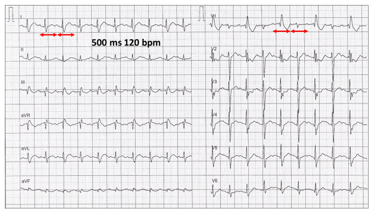
Here is an example of supraventricular tachycardia with alternating right bundle branch block.
The rate is 120 bpm at rest and the P waves regular
Here is another tachycardia example. Although the rhythm is sinus 60 bpm (red arrows), the actual rate is much faster because of atrial bigeminy (blue arrows).
This example shows interpolated atrial bigeminy with intermittent aberration rather than true right bundle branch block.
The ectopic prematurity is subtle and more obvious when magnified.
Then there is first degree AV block and alternating right and left bundle branch block. This is only rarely seen as it is transitional to complete heart block.
The next one continues to puzzle me, and I don’t have the full answer.
The resting 12-lead ECG rhythm strip lead II shows a bidirectional rhythm.
Rhythm strip: P waves are regular and almost identical. Assume sinus rhythm! The QRS complexes show alternating right (red highlight) and left (yellow highlight) bundle branch block.
Let us look at more rhythm strips.
V1 shows an interesting QRS transition pattern (red highlight).
With the right bundle branch block, the PR interval gradually increases in Wenckebach sequences, the QRS narrows (less aberration).
Anyone think of a better explanation.
I couldn’t finish without this ECG, which is frequently misdiagnosed as end diastolic ventricular bigeminy (red highlight).