To receive a free PDF copy of The Fundamentals of Electrocardiograph Interpretation by Harry Mond, subscribe to his email blog by entering your email address below.
Thank you! Your submission has been received!
Oops! Something went wrong while submitting the form.
Purchase the textbook
Purchase a hard cover or paperback copy of The Fundamentals of Electrocardiograph Interpretation by Harry Mond on Amazon.
Whilst searching this week for a “What do you think?” ECG, I reported this challenging 12-lead ECG. I changed the rhythm strip to V4, which was clearer.
I was given no clues, but to be nice, I am providing the best quality rhythm strips.
Spend some time looking at them.
What do you think? There is the prize of a smile if you get it correct!
Let us look at the rhythm strips as all the information is there.
The underlying rhythm is atrial fibrillation.
Paced beats (red highlight) with a right bundle branch block configuration (blue open circle) suggesting left ventricular or more accurately biventricular pacing. Remember, the keyword is ”configuration” as pacing is not via the normal conducting system, but rather “looks like” a right bundle branch block.
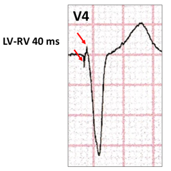
Are there two pacing spikes? Let us look at pacing in V4 (red open circle).
Two stimulus artefacts with a delay of 40 ms confirming biventricular pacing. With biventricular pacing for cardiac resynchronization therapy, the left ventricle is usually paced before the right ventricle resulting in a latency period, which allows both stimulus artefacts to be identified. In our case, the latency is LV-RV 40 ms.
The longer the programmed latency, the wider apart the two stimulus artefacts.
Even with no latency (LV-RV 0ms) two stimulus artefacts are often seen with some difficulty and the appearance is usually a left bundle branch block configuration (red highlight).With LV-RV 80 ms (yellow highlight), the two stimulus artefacts are well apart, and the appearance is now right bundle branch block configuration.
Here is another example of biventricular pacing with a right bundle branch block configuration(red highlight) and a short LV-RV latency (yellow highlight).
Our tracing also showed a number of broad QRS funny looking beats (FLBs, yellow highlight). These are usually called ventricular ectopics, but remember the underlying rhythm is atrial fibrillation, so these could be conducted native beats with a bundle branch block.
These beats are sensed by the implanted pacemaker and the next ventricular paced beat has the same cycle length (green arrow) as the paced to paced cycle length(blue arrow). Another feature is a stimulus artefact within the QRS of theseFLBs. These do not time out with the paced cycle length (blue arrow) and thus are not ventricular under-sensing. Rather, this is ventricular triggered pacing(VVT or maybe DDT), which is a program unique to biventricular pacemakers and are programmed ON as the default setting in at least two manufacturer’s products (VentricularSense Response– Medtronic Inc, Minneapolis, MN andBiV Trigger – Boston Scientific, St Paul, MN). The objective is to attempt biventricular pacing on the native QRS and attempt resynchronization, which is obviously ineffective in our case as the sensing is late and well into the depolarization.
Here is another example:
Biventricular pacing with aright bundle branch block configuration (red highlight) and two stimulus artefacts (yellow highlight). Native ventricular beats demonstrating late sensing with two stimulus artefacts, late in the QRS (blue highlight).
Let us revisit the native QRS beats (yellow highlight).
The timing cycle is very regular (red arrows), whereas the coupling interval between the paced and native beats are very variable (blue arrows). A fusion beat is present (blue highlight, with absent coupling interval).
These native beats are neither conducted from the atrium nor truly ectopics, but rather ventricular parasystole.
To summarize: Ventricular parasystole
Rare ECG finding
Independent, very slow ventricular rhythm
Sinus impulses cannot enter and reset
Sinus impulses, however, can create refractoriness of the surrounding myocardium causing an exit block.
No ventricular complexes during refractory periods (red arrow, blue glow)
Fusion beats are common depending on the timing(red stippled arrow).
Parasystole can occur with atrial fibrillation.
Atrial fibrillation with the ventricular parasystolic cycle length (1600 ms – 38 bpm) slightly faster than the narrow native conducted beats (red arrows). Thus the coupling intervals shorten (red highlight), until there is an early conducted native beat and the parasystolic focus is blocked by the refractory period (red arrow, blue glow).
Wait! We aren’t finished yet. There is another broad FLB (yellow highlight).
It is very premature and the cycle length to the next paced beat (blue arrow) is identical to the paced cycle lengths (red arrow). It also has a very early stimulus artefact (red open circle and below), as its ectopic focus must be close to the left ventricular pacing lead, particularly as it also has a right bundle branch block configuration. Once again, there are two stimulus artefacts 40 ms apart. (blue and green lines).
We now can diagnose this ECG:
Atrial fibrillation, biventricular pacing with LV-RV 40 ms latency and triggering (VVT) on nativeQRS complexes with the delay dependent on how close the native beat focus is to the pacing lead cathodes. Ventricular parasystole and a ventricular ectopic.
No tricks. Just a selection of tracings from a Holter study.Look at each one carefully, use calipers, arrows and highlight and write down your conclusions.
.png)