To receive a free PDF copy of The Fundamentals of Electrocardiograph Interpretation by Harry Mond, subscribe to his email blog by entering your email address below.
Thank you! Your submission has been received!
Oops! Something went wrong while submitting the form.
Purchase the textbook
Purchase a hard cover or paperback copy of The Fundamentals of Electrocardiograph Interpretation by Harry Mond on Amazon.
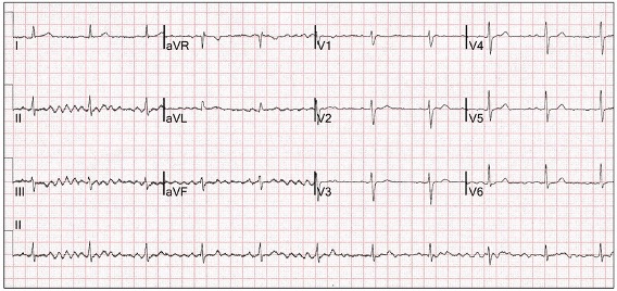
A 47-year old concert pianist presented with atypical migraine. The ECG was reported as atrial fibrillation with a controlled ventricular response.
Commenced on anticoagulants.
Now on life support in intensive care following a massive haematemesis/melaena.
What do you think?
The areas on the ECG in red highlight show sinus rhythm (red arrows). The QRS complexes in the lead II rhythm strip are regular (blue arrows) and there is modest mechanical artefact from the left leg (II, III, aVF).
This is clearly sinus rhythm and the misdiagnosis led to serious complications from anticoagulant therapy.
The reporting of atrial fibrillation on the 12-lead ECG, particularly with artefact, is the most common serious misdiagnosis with ECG reporting.There is almost always a clue to the diagnosis of sinus rhythm.
If unsure:
Carefully review each lead.
Is the ventricular rate regular?
If necessary, repeat the tracing with long rhythm strips.
The following tracings were all reported as atrial fibrillation (unless specified).
Regular sinus tachycardia (red arrows).
Interference most likely from left arm (leads I, III).
Think Parkinsonian tremor.
Regular sinus rhythm (red arrows).
Interference most likely right arm (leads I, II).
Again think Parkinsonian tremor.
Regular sinus rhythm (red arrows).
Interference most likely left arm (leads I, III).
Think Parkinsonian tremor.
This ECG was reported as atrial flutter.
Interference most likely left arm (leads I, III).
Think Parkinsonian tremor.
Poor quality ECGs with atrial ectopics are frequently diagnosed as atrial fibrillation.
Sinus rhythm (red arrows) with atrial ectopics (purple arrows).
The rhythm strip shows a regular ventricular rate (blue arrows) interrupted by premature atrial ectopics (purple arrows) and compensatory pauses.
Another example which highlights how easy it is to diagnose atrial fibrillation.
Sinus rhythm (red arrows) with premature atrial ectopics (purple arrows).
Interference most likely left leg (leads II, III).
Despite obvious artefact without a change in rate (red highlight), this was still called sinus rhythm(red arrows) with a short run of atrial fibrillation).
Sometimes artefact is subtle(yellow highlight) and suggests an irregular heart rate.
Yes, this was called atrial fibrillation, despite the obvious P waves (red arrows).
With miniscule P waves (red arrow), sinus rhythm can be difficult to diagnose.
In this case, despite the regular heart rate, the auto-interpretative reporting system diagnosed atrial fibrillation.
When it is atrial fibrillation, don’t doubt the diagnosis.
Patient with Parkinson’ disease.
The interference was recognised as such, and the diagnosis was sinus rhythm with an atrial run. It is clearly irregular and no P waves and thus atrial fibrillation.
Remember, everyone makes these errors.
Taking care with reporting, means you make them less than others.
Fusion beats are an amalgam of two competing rhythms within the same chamber, atrial or ventricular. They are not “abnormal” beats and are a lesson in timing. For fusion beats to occur, two foci must commence propagation at or near the same time and both contribute to chamber depolarization. Both must be either atrial or ventricular and depending on the contribution of each, the resultant progeny have similarities to one or both parents.