To receive a free PDF copy of The Fundamentals of Electrocardiograph Interpretation by Harry Mond, subscribe to his email blog by entering your email address below.
Thank you! Your submission has been received!
Oops! Something went wrong while submitting the form.
Purchase the textbook
Purchase a hard cover or paperback copy of The Fundamentals of Electrocardiograph Interpretation by Harry Mond on Amazon.
With ventricular pentageminy, there are unifocal ventricular ectopics with fixed coupling intervals every 5th beat with 4 sinus beats between. In this ECG the coupling intervals are not fixed.
The ventricular ectopics are late (end-diastolic) and lie within the PR intervals.
The earliest ventricular ectopic commences midway through the sinus P wave (red highlight). Another ventricular ectopic (yellow highlight) commences on the downslope of the sinus P wave, whereasa third ectopic is a fusion beat (blue highlight). The time between ventricular ectopics is fixed at 3.6 seconds.
This is most likely ventricular parasystole.
Ventricular parasystole:
Rare ECG finding
Independent, very slow ventricular ectopic focus
Sinus impulses cannot enter and reset
Sinus impulses, however, can createrefractoriness of the surrounding myocardium causing an exit block.
ECG features:
Unifocal ventricular ectopics
Variable coupling intervals
Fixed, usually very slow inter-ectopic intervals
No ventricular ectopics during the refractory periods
Fusion beats are common depending on the timing.
The diagnosis is always difficult, controversial and this even applies to cases in the literature.
To be absolutely sure, we need long strips.
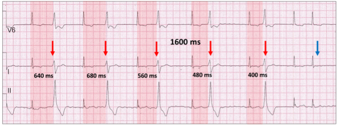
Let us look at another ECG example this time with atrial fibrillation.
The relationship or coupling intervals of the ventricular ectopic (red vertical arrow) to the previous sinus beat (red highlight) varies.
The inter-ectopic intervals are all 1600 ms apart with one in the refractory period (blue vertical arrow).
Fusion beats are an amalgam of two competing rhythms within the same chamber, atrial or ventricular. They are not “abnormal” beats and are a lesson in timing. For fusion beats to occur, two foci must commence propagation at or near the same time and both contribute to chamber depolarization. Both must be either atrial or ventricular and depending on the contribution of each, the resultant progeny have similarities to one or both parents.