To receive a free PDF copy of The Fundamentals of Electrocardiograph Interpretation by Harry Mond, subscribe to his email blog by entering your email address below.
Thank you! Your submission has been received!
Oops! Something went wrong while submitting the form.
Purchase the textbook
Purchase a hard cover or paperback copy of The Fundamentals of Electrocardiograph Interpretation by Harry Mond on Amazon.
Retrograde conduction is defined as conduction from an ectopic site to the atrium via the AV node or accessory pathway and referred to as VA conduction. II results in a P wave configuration which differs from an anterograde P wave, and in most ECG leads is inverted. The site from where the impulse originates can be the AV node, Bundle of His, distal conducting system or the body of the ventricles including ventricular pacing. On the ECG, the QRS complex usually precedes the inverted retrograde P wave but on occasion may follow it.
The two main sites for the ectopic focus that result in retrograde conduction are the AV junction and from within the ventricles.
Junctional ectopics and rhythms
With junctional rhythm, there are two scenarios of retrograde P waves.
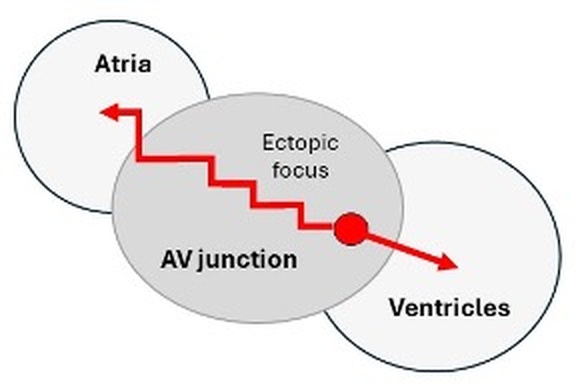
The inverted P wave follows the QRS.
The ectopic focus lies low in the AV junction, allowing the anterograde wave to depolarize the ventricles well before the retrograde wave slowly traverses the AV junction to depolarize the atria (red arrows).
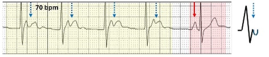
The inverted P waves follow the QRS and lies in the ST segment/T wave.
Sinus rhythm (red highlight, red arrow) and a junctional escape rhythm (yellow highlight) with the inverted P waves (blue stippled arrows, blue inverted half circle), lying within the T waves.
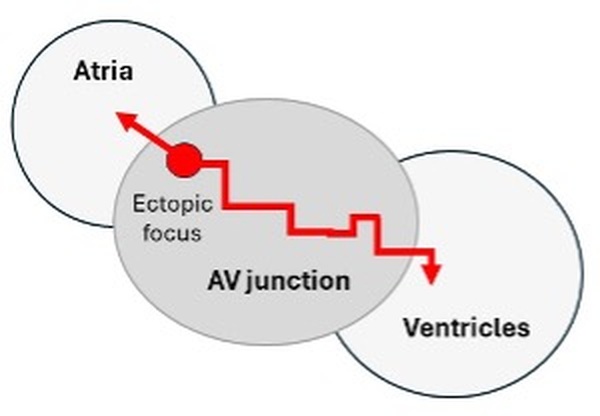
The inverted P wave precedes the QRS.
The ectopic focus lies high in the AV junction allowing the retrograde wave to rapidly depolarize the atria first, then slowly traverses the AV junction anterograde to depolarize the ventricle (red arrows).
Inverted P waves with short PR intervals.
Sinus rhythm (red highlight, red arrow) and a junctional escape rhythm (yellow highlight) with the inverted P waves prior to the QRS and the PR interval is 80 ms. (blue arrows, blue inverted half circle).
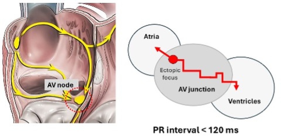
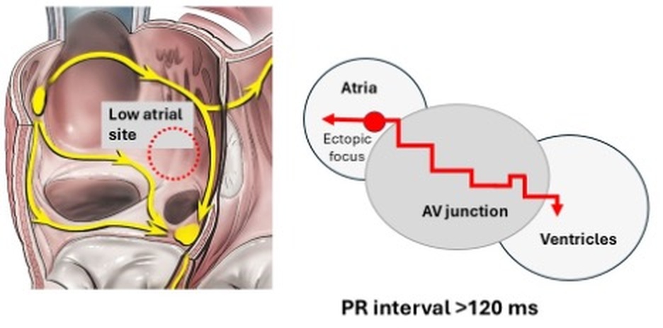
The ECG configuration with a preceding inverted P wave is similar to that seen with a low atrial rhythm. This is not surprising as a low atrial rhythm originating adjacent to or from within the triangle of Koch is very close to the AV junction. A reported rule for a junctional rhythm (red stippled open circle) is the PR interval should be <120 ms.
With a low atrial site (red open stippled circle), conduction is delayed and > 120 ms.
The electrophysiological properties of the AV junction vary and therefore setting a time limit is of little value. Therefore, all these ECG appearances irrespective of the PR interval can be called ectopic atrial, low atrial or junctional ectopics or rhythms.
Ventricular ectopy
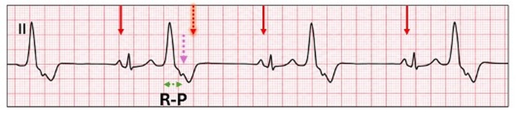
With ventricular ectopy and VA conduction, the inverted P wave lies in the ST segment or T wave of the ectopic with the timing of the conduction being from the onset of the QRS to the commencement of the P wave or where it is best recognised. The timing is referred to as the RP interval. It is not always easy to recognise these P waves, which are frequently mistaken for non-conducted sinus P wave.
Ventricular bigeminy with retrograde P waves (purple stippled arrow). The inverted P wave is earlier than the next scheduled sinus P wave (shadowed stippled red arrow). Type image caption here (optional)
When unsure if there is retrograde conduction or a non-conducted sinus P wave, measurement of the compensatory pause will provide the answer.
In summary:
Ventricular full (complete) compensatory pause
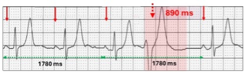
A full compensatory pause necessitates that there is no retrograde conduction to the atrium and thus the sinus cycle is not reset by the ventricular ectopic. The next scheduled sinus P wave cannot conduct to the ventricle as the pathway is still refractory. To determine the compensatory pause, measure two sinus cycles, one with the embedded ventricular ectopic and two preceding or subsequent sinus cycles.
Ventricular ectopic (red highlight) with a full compensatory pause. The ventricular ectopic does not reset the sinus cycle and thus the next scheduled P wave lies within the ventricular ectopic (red stippled arrow). In timing, two sinus cycles with the embedded ventricular ectopic is the same as two sinus cycles following the atrial ectopic (1780 ms). The compensatory pause covers the “full” sinus cycle (890 ms).
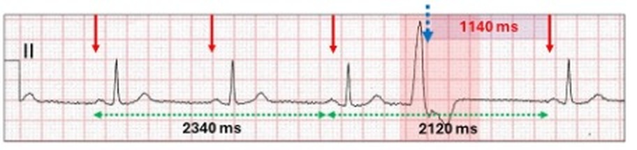
The sinus node is reset by retrograde conduction to the atrium and because it is earlier than the next scheduled sinus beat, the ventricular compensatory pause is partial. It is best seen when comparing two cycles, one with the embedded ventricular ectopic and one without.
Ventricular ectopic (red highlight) having a partial compensatory pause. Because the retrograde P wave (blue stippled arrow) resets the sinus node, the timing of two sinus complexes with the embedded atrial ectopic is shorter (2120 ms) than the two sinus cycles preceding the ventricular ectopic (2340 ms). The timing of the compensatory pause is from the retrograde P wave to the next scheduled sinus beat (1140 ms).
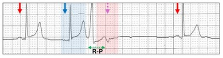
When retrograde conduction is delayed, there is a long RP interval (first degree VA block) and the inverted P wave may emerge from the ectopic T wave.
Sinus rhythm (red arrows) with a possible junctional ectopic (blue highlight, blue arrow) followed by a narrow QRS ventricular or fascicular ectopic with retrograde conduction (pink stippled arrow) and a prolonged R-P interval. The earlier junctional ectopic may result in a refractory fast pathway, allowing the retrograde conduction from the ventricular ectopic to conduct via the slow pathway, resulting in a prolonged RP interval.
Ventricular arrhythmias
P waves are usually difficult to identify with ventricular tachyarrhythmias. If the rhythm is slow, then the non-conducted sinus P wave can occasionally be recognised and with pauses, AV conduction may be present with a sinus generated QRS.
Short runs of ventricular tachycardia with a background of conducted (solid red arrow, red highlight) and non-conducted (stippled red arrow) sinus P waves.
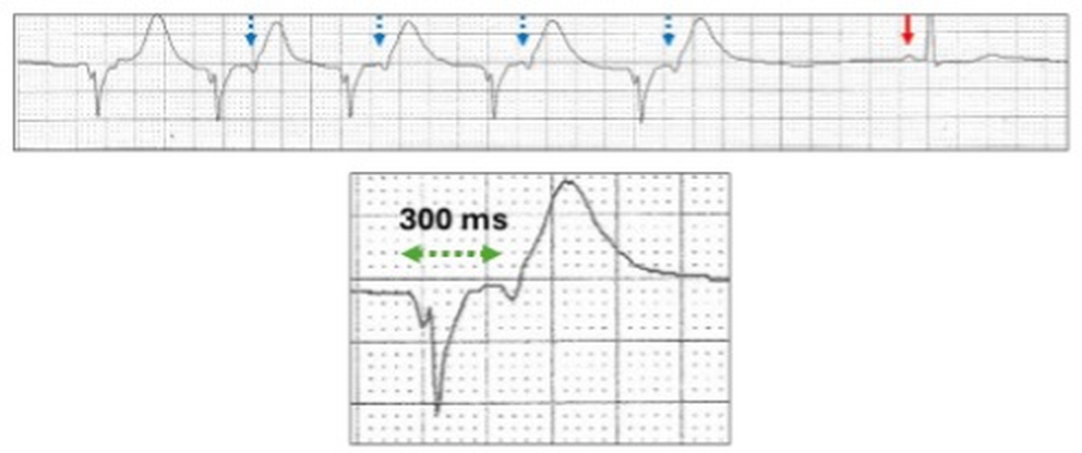
With a slow sinus rhythm, 1:1 VA conduction can occur.
Idioventricular run with obvious 1:1 retrograde P waves at the end of the ST segment. The RP interval is long (300 ms) and this is called first degree AV block.
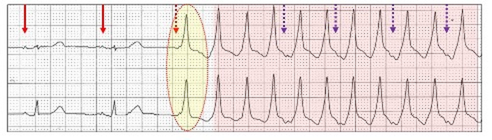
VA conduction however is very fragile and blocks occur with faster rates.
Sinus rhythm (red arrows) with a run of ventricular tachycardia and 2:1 VA block (purple stippled arrows). The tachycardia commences with a fusion beat (yellow oval).
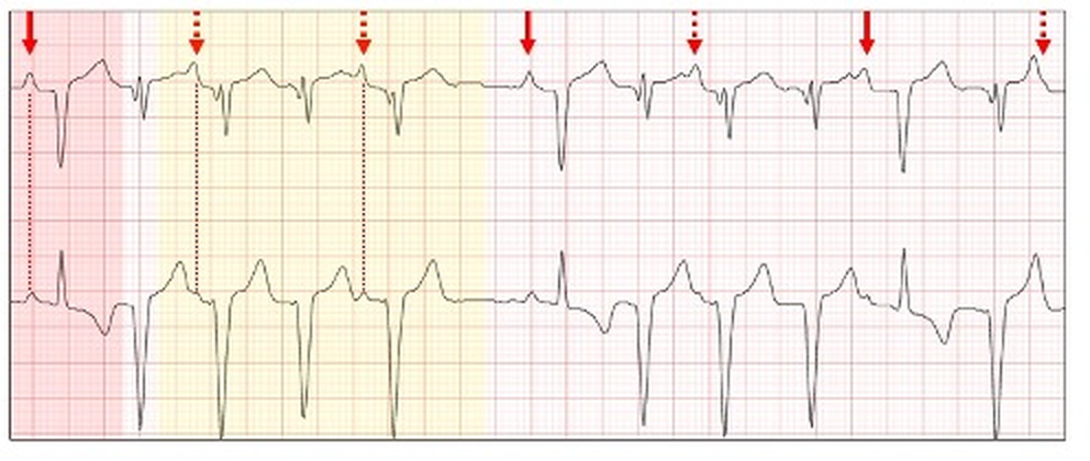
More complicated VA blocks can occur usually with slow ventricular rhythms.
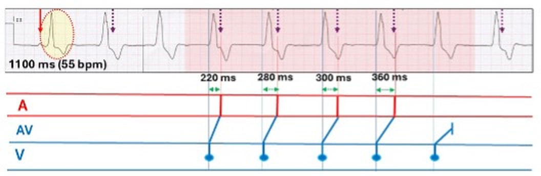
ECG strip and laddergram. The rhythm commences as sinus (red arrow) with ventricular fusion (yellow oval) followed by a run of idioventricular rhythm, 55 bpm (red highlight) with poorly seen retrograde P waves (purple stippled arrows). The VA conduction times (green stippled arrows) gradually increase from 220 ms to 360 ms before VA conduction blocks and the next sequence starts. This is a 5:4 VA Wenckebach sequence.
There is controversy as to whether anterograde and retrograde conduction use the same AV pathways. Although dual AV nodal pathway physiology may play a role, some authorities believe that within a pathway, there are separate anterograde and retrograde channels. This might explain why bidirectional AV conduction may not have the same timing. When AV nodal disease is present, it becomes more complicated. For instance, with complete AV block, there is no anterograde conduction but in about 10% of cases, retrograde conduction is possible.
VVI pacing in a patient with sinus bradycardia and complete AV block. There is retrograde conduction to the atrium with each paced beat (red oval).
Retrograde conduction is defined as conduction from an ectopic site to the atrium via the AV node or accessory pathway and referred to as VA conduction. II results in a P wave configuration which differs from an anterograde P wave, and in most ECG leads is inverted. The site from where the impulse originates can be the AV node, Bundle of His, distal conducting system or the body of the ventricles including ventricular pacing. On the ECG, the QRS complex usually precedes the inverted retrograde P wave but on occasion may follow it.





.png)





