To receive a free PDF copy of The Fundamentals of Electrocardiograph Interpretation by Harry Mond, subscribe to his email blog by entering your email address below.
Thank you! Your submission has been received!
Oops! Something went wrong while submitting the form.
Purchase the textbook
Purchase a hard cover or paperback copy of The Fundamentals of Electrocardiograph Interpretation by Harry Mond on Amazon.
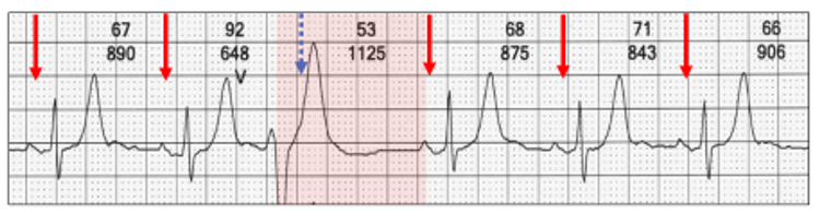
There is sinus rhythm (arrows) and a ventricular ectopic (red highlight).
Embedded in the terminal portion of the T wave is a sinus P wave (blue arrow). Because of the timing of ventricular ectopic, the P wave falls within the refractory period of the ventricle and therefore is not conducted.
With ventricular ectopics, a sinus P wave is usually concealed (blue highlight) within the ectopic and obviously does not conduct, unlike the other P waves (red arrows).
A poorly understood feature of ventricular ectopics is that the non-conducted sinus P wave (blue arrow) may not be concealed within the ectopic QRS (yellow highlight).
With appropriate timing (including sinus rate, ectopic prematurity) as well as QRS width and shape, the sinus P wave may emerge from the tail of the QRS or even the T wave.
Left ECG: The sinus rate is ~60 bpm (blue highlight) and the non-conducted sinus P wave (blue arrow) lies at the end of the T wave.
Right ECG: The sinus rate is ~50 bpm (blue highlight) and the non-conducted sinus P wave is later (blue arrow), but still within the refractory period of the ectopic. This mimics second degree AV block. I call this pseudo AV block and remember it is physiologic and dependent on the timing.
Fusion beats are an amalgam of two competing rhythms within the same chamber, atrial or ventricular. They are not “abnormal” beats and are a lesson in timing. For fusion beats to occur, two foci must commence propagation at or near the same time and both contribute to chamber depolarization. Both must be either atrial or ventricular and depending on the contribution of each, the resultant progeny have similarities to one or both parents.