To receive a free PDF copy of The Fundamentals of Electrocardiograph Interpretation by Harry Mond, subscribe to his email blog by entering your email address below.
Thank you! Your submission has been received!
Oops! Something went wrong while submitting the form.
Purchase the textbook
Purchase a hard cover or paperback copy of The Fundamentals of Electrocardiograph Interpretation by Harry Mond on Amazon.
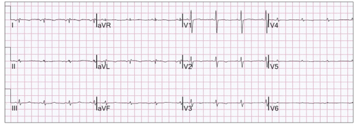
I was shown this ECG as a good example of dextrocardia.
What do you think?
Dextrocardia has characteristic ECG findings:
Limb leads:Identical to reversed arm leads:
Lead I is flipped.
aVR and aVL are reversed.
Leads II and III are reversed – usually not helpful in the diagnosis.
Chest leads: When the chest leads are placed in the traditional manner, leads V1 and V2 are reversed, and the remaining V leads are moving away from the heart with absentR waves and ever decreasing QRS size.
To visualize the normal ECG, the leads should be placed across the right chest as shown.
Here is another example of dextrocardia:
In our ECG, the features of reversed arm leads are present and consistent with dextrocardia.
Lead I is flipped.
aVR and aVL are reversed
Leads II and II are reversed.
What about the chest leads?
The chest leads have all been reversed, so that V1 is in the V6 position and V6 is in the V1 position.
So reviewing our ECG:
The limb leads are reversed (consistent with dextrocardia)
The chest leads are reversed (not consistent with dextrocardia).
I call this “The Full Hand” in that both the arm and chest leads are reversed.
Although rare, I find this more common than dextrocardia.
Reversal in only the limb or chest leads is not uncommon.
Limb lead reversal (yellow highlight):
Chest lead reversal (yellow highlight):
I love to finish with a doozy! I reported this as dextrocardia.
I was told the patient’s heart had normal orientation.
I found an ECG done a year before:
This is :
Reversed limb leads
Chest leads on the right side.
I suspect the person recording the ECG saw poor progression of R waves across the chest leads and mistook this for possible dextrocardia, hence reversing the arm leads and chest leads to the right.
Fusion beats are an amalgam of two competing rhythms within the same chamber, atrial or ventricular. They are not “abnormal” beats and are a lesson in timing. For fusion beats to occur, two foci must commence propagation at or near the same time and both contribute to chamber depolarization. Both must be either atrial or ventricular and depending on the contribution of each, the resultant progeny have similarities to one or both parents.