To receive a free PDF copy of The Fundamentals of Electrocardiograph Interpretation by Harry Mond, subscribe to his email blog by entering your email address below.
Thank you! Your submission has been received!
Oops! Something went wrong while submitting the form.
Purchase the textbook
Purchase a hard cover or paperback copy of The Fundamentals of Electrocardiograph Interpretation by Harry Mond on Amazon.
Well at the end of this one you will have a headache, so take two aspirin now!
Our last ECG topic was reversed arm leads and dextrocardia.
This topic also looks at twisted leads, but this time involving all limbs.
I have discussed this before (Fun with ECGs 28), but I would like to take it further.
Let us twist the arm and leg leads with “leg leads on arms” and “arm leads on legs”.
To summarize:
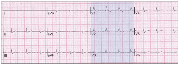
Major footprint: Lead I is now on the feet well away from the heart and thus isoelectric (red highlight).
There are three minor footprints:
All limb leads are often attenuated
Leads II and III and aVF are near identical (yellow highlight)
Leads aVR and aVF are near identical (green highlight)
Here it is again (no limb lead attenuation):
Because the limb leads are reversed superior/inferior, it is not surprising that the P waves may also be reversed in leads II, III and aVF:
A right bundle branch block (blue highlight) makes no difference.
Sometimes reversed limb leads are overlooked because other abnormalities overwhelm you!
There is a prolonged QT interval (blue highlight).
Reversed limb leads are not always as easy to diagnose as these ECGs suggest.
There is growing popularity in placing the leg lead electrodes on the abdomen.
This makes no difference as far as conventional ECG are concerned.
Does this affect reversed limb leads? RA and LA leads are now near the heart.
The QRS emerges in lead I.
The clues are there, but very easy to miss.
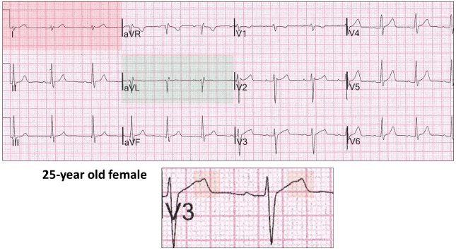
Here is an example which is probably a normal variant and not reversed limb leads.
Axis to the right (red highlight). We have previously said that aVL is negative (green highlight) in this situation (Fun with ECGs 41). This mimics reversed limb leads.
Note the notched T wave in V3. We will discuss this further one day.
Now take two aspirin as we will look further with twisted leads next time!
Remember, keep your eyes open when reporting ECGs!
Fusion beats are an amalgam of two competing rhythms within the same chamber, atrial or ventricular. They are not “abnormal” beats and are a lesson in timing. For fusion beats to occur, two foci must commence propagation at or near the same time and both contribute to chamber depolarization. Both must be either atrial or ventricular and depending on the contribution of each, the resultant progeny have similarities to one or both parents.